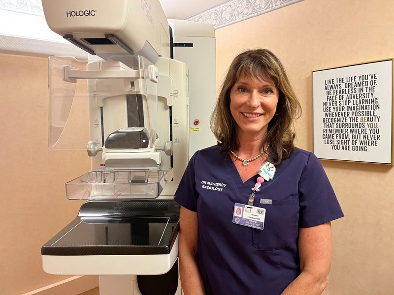
Jennifer Mayberry
“Screening for breast cancer is one of the most important things a woman can do for her health,” said Jennifer Mayberry, MD, who specializes in in diagnostic radiology and is the Breast Imaging Director at Tri-City Medical Center. “If it is found early, breast cancer is easier to treat, the need for chemotherapy may be eliminated and survival rates are higher. At Tri-City, we use three different types of non-invasive breast imaging tests – mammogram, ultrasound and MRI – depending on the individual’s situation.”
A mammogram is the most common, routine screening test for breast cancer. The American College of Radiology and Society of Breast Imaging recommends annual mammography beginning at age 40, or younger if the patient’s mother had breast cancer in her 40s.
A two-dimensional (2D) digital mammogram is the standard used to look for signs of breast cancer when there are no symptoms. Low-dose x-rays are taken of the breast, from two different angles (top/bottom and side-to-side) and stored digitally. If symptoms such as a lump, pain or nipple discharge are present, or something suspicious appears on a screening mammogram, then a healthcare provider will order a diagnostic mammogram that provides more specifically directed images.
During digital breast tomosynthesis (DBT), also called 3D mammography, multiple low-dose x-rays are taken of the breast from different angles as the machine moves around the breast. A computer compiles the images together into a series of thin slices creating a much clearer image than 2D.
“DBT allows us to visualize that breast tissue more thoroughly,” said Dr. Mayberry. “Think of it like a book. Lying on the table, the book only shows two views with all of the pages overlapping. But by standing the book up on its spine, we can flip through multiple pages and see more of the words and pictures. This procedure is especially helpful for women with dense breasts, which make it more difficult to see masses or other abnormalities.”
According to the American Cancer Society, women with dense breast tissue have a higher risk of developing breast cancer than those who do not. The US Food and Drug Administration (FDA) issued a rule in March 2023 stating that mammogram reports sent to patients must include breast density describing it as either “not dense” or “dense;” all states must comply by September 2024. TCMC already includes this information in patient mammogram reports.
“Another way we can look at the breast is by using ultrasound,” continued Dr. Mayberry. “Sound waves allow us to see the tissue and its properties in real time, and differentiate between fat, a fluid-filled cyst or a solid mass. We can also use ultrasound to check if the lymph nodes under the arm (axilla) are normal or abnormal.”
Instead of sound waves, breast magnetic resonance imaging (MRI) uses radio waves, magnets and a computer to create detailed, cross-sectional pictures of the breast’s inner soft tissue. For this procedure, contrast dye must be injected into a person’s vein through an IV to make any abnormalities easier to see in the pictures. This is the most sensitive and specific of the three tests and may be used to screen women who have a higher risk of breast cancer due to family or personal history of the disease, gene mutations, dense breast or older age.
“Since we have recently added a 3Tesla MRI suite at Tri-City, we are also going to be performing a newer technique called abbreviated breast MRI or fast breast MRI by the beginning of next year,” said Dr. Mayberry. “It’s similar to Standard 2T breast MRI but fewer images are taken over a shorter timeframe. It’s an additional tool for high-risk patients and is currently being studied as a possible screening test for women with dense breasts.”
“Tri-City is recognized as a Breast Imaging Center of Excellence by the American College of Radiology (ACR),” added Dr. Mayberry. “The Breast Imaging Center at Tri-City meets the Federal standards 100% as set forth by the Mammography Quality Standards Act (MQSA) and enforced through accreditation by the ACR. When it comes to breast cancer imaging, we offer the expertise and advanced technologies needed to detect breast cancer when it is small and has not spread. These two important factors and early detection play a big part in determining a person’s prognosis with this disease.
“We want all of our employees to be aware that we are available to provide breast imaging services before and after their scheduled work day, Monday – Thursday. It is a quick and easy test to ensure the continued health of our Tri-City family.”


